|
Erythema Nodosum (EN)
Erythema nodosum (EN) is an inflammation of the subcutaneous fat characterized by
painful, red swellings over the shins. The eruption may also involve the extensor aspects of the
thighs and forearms. The condition is believed to be a delayed hypersensitivity reaction and occurs
most often in women during their
reproductive years [2, 3].
 Erythema nodosum migrans, subacute nodular migratory panniculitis, and chronic
erythema nodosum, are considered to be variants of the same disease [1]. Erythema nodosum migrans, subacute nodular migratory panniculitis, and chronic
erythema nodosum, are considered to be variants of the same disease [1].
Excisional biopsy of the lesions in the early stages of EN shows a perivascular infiltrate of neutrophils in the interlobular septa
that is sub-sequently replaced by lymphocytes, and a granulomatous infiltrate
with giant cells. Small histiocytes, radially placed around a central cleft, Miescher's radial granulomas,
are a characteristic finding.
EN
may be associated with a variety of systemic diseases or medications.
However, an identifiable cause is not found in about 50% of patients. The
most common identifiable causes of EN are streptococcal infection and sarcoidosis [1-5].
Conditions Associated with Erythema Nodosum
Common [1 -16]
|
Infections
|
|
|
Streptococcal phyaryngitis
Tuberculosis
Coccidioidomycosis
Yersinia
Histoplasmosis
|
Sarcoidosis
Drugs |
|
|
Sulfa drugs
Oral
contraceptives
Amoxicillin |
Inflammatory bowel disease
Behçet disease
Pregnancy (most often during the first half of
pregnancy [12]) |
Less Common [1-4,16]
|
Infections
|
| |
Campylobacter
Rickettsiae
Salmonella
Psittacosis
Syphilis
Amoebiasis Giardiasis
Herpes simplex virus
Mycoplasma
Epstein-Barr virusHepatitis B and C virusesHuman immunodeficiency virus
ToxoplasmoisCat scratch disease (Bartonella) |
|
Malignancies
|
| |
Leukemia
Hodgkin’s disease
|
Evaluation
Initial evaluation shouldincludes:
- Throat culture>
- Antistreptolysin-O (ASO) titer
- CBC
-
Erythrocyte sedimentation rate , ESR
- Intradermal skin tests for tuberculosis
and coccidioidomycosis
- Chest film to evaluate for hilar adenopathy.
- In patients with GI symptoms obtain stool culture to include Yersinia
enterocolitica (consider serological testing also).
- Consultation with a dermatologist and/or internist for evaluation of
underlying cause of EN may be helpful.
If a biopsy is necessary to confirm the diagnosis , deep skin incisional
biopsies are required to sample the subcutaneous tissue adequately.
Other conditions to consider in the differential diagnosis include
alpha-1 antitrypsin deficiency panniculitis, lupus panniculitis, cutaneous
polyarteritis nodosa,
superficial thrombophlebitis , and erythema induratum.
Clinical Course
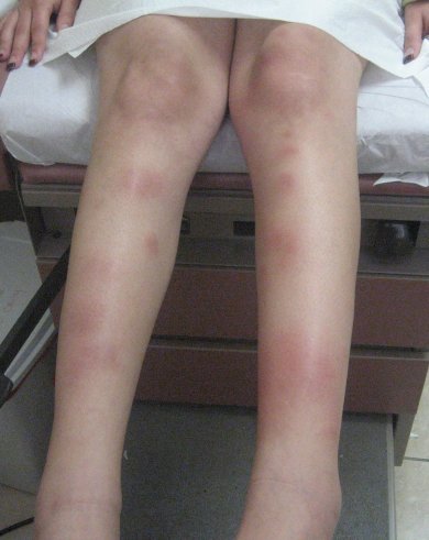
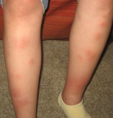
EN typically presents as multiple, tender, bright red, 1 to 20 cm in diameter, poorly
demarcated nodules on the shins (usually bilateral). The eruption may also involve the extensor aspects of the thighs and forearms.
Over the course of 2 weeks the the lesions change in color to a bluish hue, and then fade to resemble a bruise. Ulceration and scarring does not normally occur.
 Ankle swelling and joint pain are common and usually resolve within a few weeks.
However, joint pain has been reported to persist for up to 2 years [4]. Pulmonary hilar
adenopathy may also develop.
Ankle swelling and joint pain are common and usually resolve within a few weeks.
However, joint pain has been reported to persist for up to 2 years [4]. Pulmonary hilar
adenopathy may also develop.
Prodromal symptoms of fatigue and malaise or upper respiratory
tract infection often precede the eruption by 1 to 3 weeks. In a study by Metz
and coworkers patients with fever, leukocytosis, elevated CRP level ( 6 X
upper limits),
accelerated ESR, presence of cough, sore throat, diarrhea, arthritis, and
pulmonary pathology were more likely to have EN secondary to an
identifiable cause [2].
EN is characteristically self limited and resolves on its own in 3 to 6 weeks.
Adverse effects upon the course of pregnancy or fetal outcome would not be
expected in idiopathic cases [8]. Underlying causes of EN that might result in
increased morbidity or mortality in the mother or fetus should be excluded.
Treatment [1,12,17,18]
Treatment of EN is aimed at the underlying disease or removal of the offending drug
when identified. Otherwise treatment during pregnancy is supportive and includes cool wet
compresses, elevation, and rest. Acetaminophen may be used for pain relief,
but nonsteroidal anti-inflammatory drugs (NSAIDs) are avoided because of their potential to
cause oligohydramnios and constriction of the ductus arteriosus.
Systemic steroids at a dosage of 1 mg per kg daily may be used for more
persistent complaints if underlying infection,
risk of sepsis, and malignancy have been excluded.
Oral prednisone at a dosage of 60 mg every morning is a typical dose.
REFERENCES
1. Schwartz RA, Nervi SJ.Erythema nodosum: a sign of systemic disease.
Am Fam Physician. 2007 Mar 1;75(5):695-700.PMID: 17375516
2.Mert A, Ozaras R, Tabak F, Pekmezci S, Demirkesen C, Ozturk R. Erythema nodosum:
an experience of 10 years. Scand J Infect Dis. 2004;36(6-7):424-7nPMID: 15307561
3.Cribier B, et al Erythema nodosum and associated diseases. A study of 129 cases. Int J Dermatol. 1998 Sep;37(9):667-72. PMID: 9762816
4. Habif, Thomas "Erythma Nodosum" Clinical Dermatology A color Guide to Diagnosis
and Therapy. , 5th ed. Ed. Thomas Habif, MD. New York: Mosby, 2010. 720-721
5. Richards WE, et al. Erythema nodosum associated with streptococcal
infection in pregnancy.Infect Dis Obstet Gynecol. 1995;3(4):166-8.PMID: 18476042
6. Arsura EL, Erythema nodosum in pregnant patients with coccidioidomycosis.Clin Infect Dis. 1998 Nov;27(5):1201-3.PMID: 9827269
7.Rosales Estrada G, et al. Erythema nodosum and pregnancy. Report of a case]Ginecol Obstet Mex. 1991 Jun;59:181-3. PMID: 1937121
8. Langer R, et al. Erythema nodosum associated with pregnancy. Case reports.Eur J Obstet Gynecol Reprod Biol. 1979 Dec;9(6):399-401.PMID: 264104
9. Coaccioli S, Onset of erythema nodosum during pregnancy: a case report.Clin Exp Obstet Gynecol. 1998;25(1-2):40-1. PMID: 9743879
10. Bombardieri S, Erythema nodosum associated with pregnancy and oral contraceptives. Br Med J. 1977 Jun 11;1(6075):1509-10. PMID: 871635
11. Daw E.Recurrent erythema nodosum of pregnancy.Br Med J. 1971 Apr 3;2(5752):44. PMID: 5102493
12. Bartelsmeyer JA, Petrie RH. Erythema nodosum, estrogens, and pregnancy.Clin Obstet Gynecol. 1990 Dec;33(4):777-81.
PMID: 2289344
13. Sams W.M., Winkelmann R.K.: The association of erythema nodosum with
ulcerative colitis. South Med J 61. 676-679.1968PMID: 5664514
14. Mert A, Ozaras R, Tabak F, Ozturk R. Primary tuberculosis cases presenting with erythema nodosum. J Dermatol. Jan 2004;31(1):66-8.PMID: 14739509
15. Farhi D, Cosnes J, Zizi N, et al. Significance of erythema nodosum and pyoderma gangrenosum in inflammatory bowel diseases: a cohort study of 2402 patients. Medicine (Baltimore). Sep 2008;87(5):281-93.
PMID: 18794711
16. Sullivan R, Clowers-Webb H, Davis MD. Erythema nodosum: a presenting sign of acute myelogenous
leukemia. Cutis. Aug 2005;76(2):114-6PMID:16209157
17. Myers SA and Murray JC. Antecedent Skin Conditions In:
Principles and Practice of Medical Therapy in Pregnancy
3rd ed,Gleicher N. et al Ed Norwalk, CT: Appleton Lange 1998, pp 1365
18. Jones SV and Black M. Effect of Pregnancy on Other Skin Disorders. In:
Obstetric and Gynecologic Dermatology 2nd ed. Black M et al Ed. St. Louis, Mo: Mosby 2002 pp 55- 56 |
