The Neuron
A neuron is an electrically excitable cells the receives and sends information allowing communication within the body. The neuron can be broken down into three basic parts:
- Dendrites receive incoming information
- Soma (cell body) is the biosynthetic portion of the cell and integrates that incoming information
- Axon sends electrical impulse from the cell body to the axon terminals to communicate with other cells.
Figure 1: Basic structure of a neuron
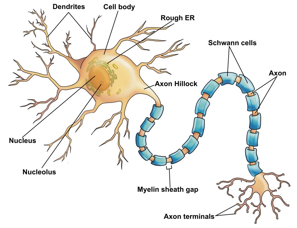
Figure 2: Soma and neuronal processes of a neuron with and without illustration overlay
Due to the specialization of neurons, there are some features not seen in other cells. Nissl bodies or chromatophilic substance are visible in neuron cell bodies under the microscope, and give the cytoplasm a granular appearance. Nissl bodies consist of ribosomes and rough endoplasmic reticulum allowing for large amounts of protein synthesis. Action potentials are the electrical impulses that neurons send from the cell body along the axon to the axon terminal. The incoming signals are integrated and the outgoing action potential originates in an area called the axon hillock, where the axon emerges from the cell body.
The axon ends at an axon terminal where it typically synapses with another neuron or muscle cell. A synapse is a site of transmission between the neuron and other cell where communication occurs. When the electrical impulse from the axon reaches the terminals, calcium influx triggers the release of a neurotransmitter from vesicles in the axon terminus. The neurotransmitter diffuses through a small gap between the axon terminal and the other cell called a synaptic cleft. Most neuronal synapses between two neurons occur between the axon terminal of one neuron and the dendrites (axodendritic) or cell body (axosomatic) of another neuron. When a axon terminal synapses with a skeletal muscle cell, it is referred to as the neuromuscular junction and the neurotransmitter released is acetylcholine (ACh).
Figure 3: The neuromuscular junction with and without illustration overlay
The axons of neurons are often myelinated. That is they are insulated by layers of myelin (protective layer of lipids and proteins). In the peripheral nervous system, myelination occurs when a Schwann cell wraps itself around the axon multiple times resulting in several layers of myelin. The cytoplasm and nucleus of the Schwann cells gets pushed to the outer layer called the outer collar of perinuclear cytoplasm or neurilemma. Along the axon there are gaps in the myelin sheath called myelin sheath gaps or nodes of Ranvier. These act to facilitate the electrical impulses along the axon. A thin layer of connective tissue surrounds the Schwann cell-axon unit called the endoneurium.
This will be discussed more in the next chapter: Central and Peripheral Nervous Systems
